Types of biopsy
- Thoracoscopic (VATS) pleural biopsy
- Laparoscopic peritoneal biopsy
- CT-guided needle biopsy
- Open surgical biopsy
Free, confidential case reviews for patients and families in all 50 states
Diagnosis & treatment
What the tests mean, why a tissue biopsy matters more than fluid cytology, and which documents a case review needs.
Confirming mesothelioma takes more than a scan. Doctors need to see the cells themselves, and the way those cells are collected and examined decides how confident the diagnosis is. This guide walks through the difference between fluid testing and a tissue biopsy, what the pathology report is actually telling you, and why a second look by a specialist pathologist is so often worthwhile. It also covers which details from these tests matter later, when treatment and any claim are being planned.
CT, PET and MRI scans are excellent at showing where something abnormal is happening, such as fluid, thickening of a lining or enlarged lymph nodes. What they cannot do is say which kind of cells are causing it. Pleural thickening can come from old scarring, infection, benign asbestos-related plaques or several different cancers. Abdominal fluid can come from liver disease, heart failure or ovarian cancer. For that reason, a report that says a scan is consistent with mesothelioma is a strong signal to keep investigating, not a final answer. The images do guide the next step, though, by showing doctors the safest and most useful place to collect a sample and whether the disease appears limited enough to make surgery worth discussing later.
When fluid has collected around a lung or in the abdomen, draining it is often the first procedure. The fluid is spun down and the cells are examined, a test called cytology. Sometimes cytology clearly shows malignant cells and even suggests mesothelioma, but it often misses the diagnosis or cannot tell mesothelioma apart from other cancers or from irritated, reactive lining cells. Mesothelioma cells can also be sparse in fluid. That is why a negative or unclear fluid result does not rule the disease out, especially when fluid keeps returning. Many centers now run additional stains on cells from fluid, which can strengthen the result. Still, most specialists prefer a tissue sample when it can be obtained safely, because it shows how the cells are behaving within the surrounding tissue.
The type of biopsy depends on where the disease is. For the chest, a surgeon often performs a thoracoscopy, also called VATS, passing a camera and instruments through small cuts between the ribs to remove pieces of the pleura. The same procedure can drain fluid and sometimes seal the space to stop it returning. For the abdomen, a laparoscopy works the same way through small cuts in the belly. A CT-guided needle biopsy can reach some areas without surgery, although the samples are smaller. Open surgical biopsies are used less often. Larger, well-chosen samples help the pathologist see whether tumor cells are invading deeper tissue, which is one of the clearest signs separating mesothelioma from harmless reactive changes.
A pathology report can feel like a foreign language, but a few parts are worth finding. The diagnosis line states the conclusion, such as malignant mesothelioma, and should name the cell type: epithelioid, sarcomatoid or biphasic. A section on immunohistochemistry lists the stains used. Mesothelioma usually stains positive for markers such as calretinin, WT-1 and D2-40, while stains that point to lung or other cancers are negative. Some reports mention BAP1 loss or other tests that help distinguish cancer from benign lining cells. Look for the date the report was finalized, the name of the pathologist and the facility. If the wording is hedged, for example suspicious for or cannot exclude, ask your doctor what further testing is planned.
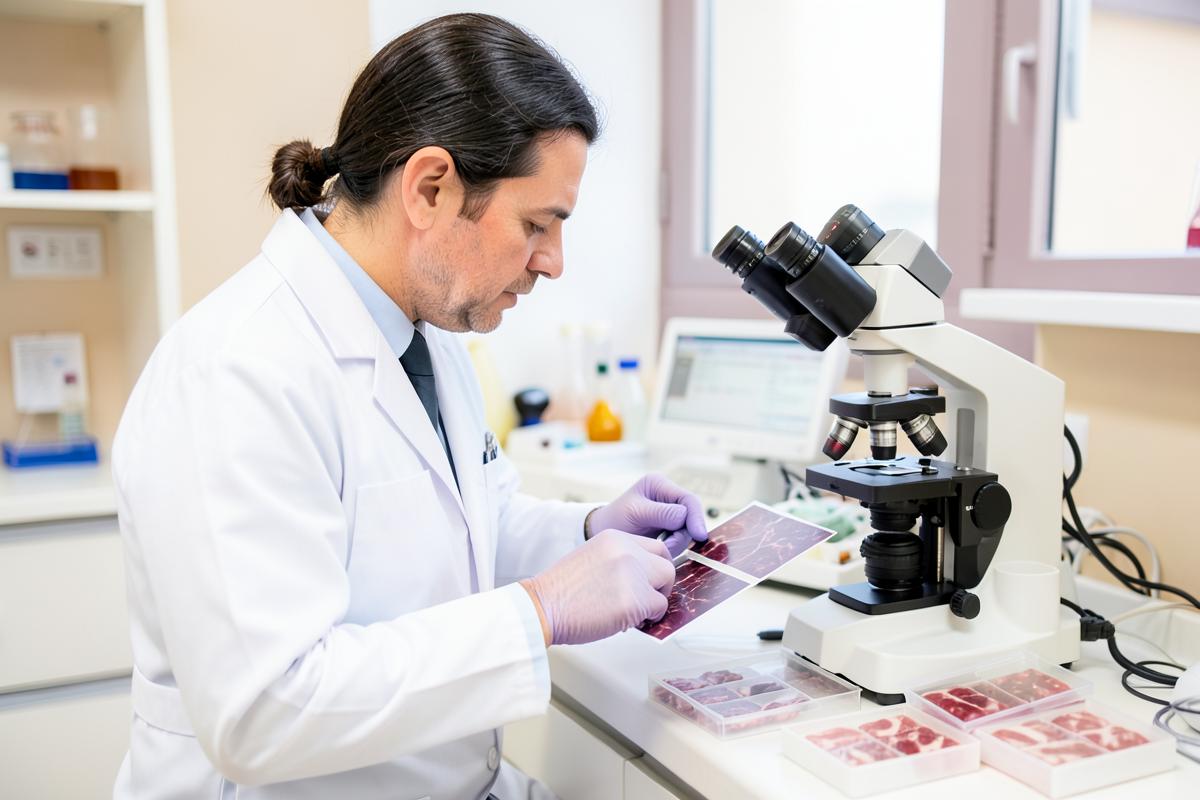
Mesothelioma is rare, so many general pathologists see only a handful of cases in their careers. Well-differentiated or early tumors can resemble reactive changes, and some mesotheliomas look like lung, ovarian or other cancers that have spread to a lining. Mistakes happen in both directions. A second review at a center that sees mesothelioma regularly can confirm the diagnosis, clarify the cell type and occasionally change the diagnosis altogether. This matters for treatment, since cell type strongly influences whether surgery is offered, and for planning more broadly. Arranging a review usually means asking the original hospital to send slides or tissue blocks to the second center. Insurance often covers it, and it rarely delays treatment by long if requested early.
Quick reference
Useful to print or bring to an appointment.
Further reading
Practical answers from our resource library.
Questions
Related
Free, confidential case review for patients and families in all 50 states.