Diagnosis
PET-CT: When It Is Used
PET-CT highlights metabolically active tissue and helps stage disease before surgery
- Topic
- Diagnosis & symptoms
- Reading time
- 3 min
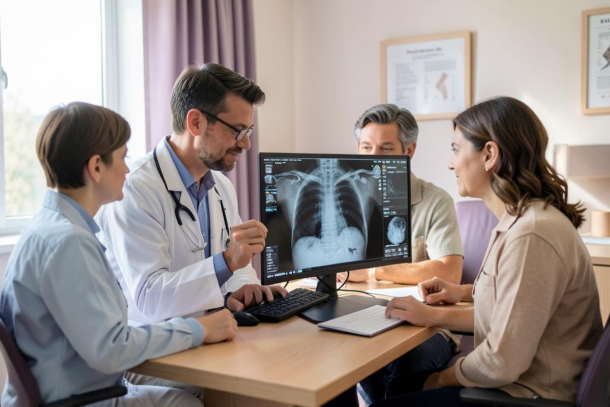
A PET-CT scan pairs a CT with a small dose of radioactive sugar. Active cancer cells tend to use more sugar, so they show up brighter on the images. For mesothelioma, PET-CT is mainly a staging tool. It helps doctors see whether disease has reached lymph nodes or distant sites, which strongly affects whether major surgery makes sense.
How the scan day works
You are usually asked not to eat for several hours beforehand, and people with diabetes get specific instructions about blood sugar and medication. After an injection of the tracer, you rest quietly for a period while it spreads through the body. Talking and moving are kept to a minimum because muscle activity can affect the images. The scan itself takes roughly twenty to thirty minutes on a table that moves through the machine. Drinking fluids afterward helps clear the tracer. Staff may suggest limiting close contact with small children or pregnant people for a short time.
Why surgeons rely on it
Before an operation, surgeons need to know whether disease is limited to one side of the chest. PET-CT can reveal active lymph nodes in the middle of the chest, spread across the diaphragm, or distant sites that a regular CT might miss. If it shows spread that surgery could not address, the team may recommend other treatment first. PET-CT findings are often confirmed with further tests before ruling surgery out, because a bright spot is a clue rather than proof. Some surgeons also use it to choose the best lymph node to sample. When PET results are unclear, a needle or scope procedure may be used to check nodes directly.
Understanding bright spots and false alarms
Inflammation and infection also use extra sugar, so they can light up too. This is a particular issue after talc pleurodesis, which causes lasting inflammation in the lining that can look active on PET for a long time. Recent procedures, healing wounds and even muscle strain can cause similar effects. That is why reports describe uptake values and possible explanations rather than simply declaring cancer. If you had pleurodesis, make sure the team reading your scan knows the date. Ask your oncologist how much weight the PET findings carry in your plan.
What to do next
- 1
Follow fasting and diabetes instructions exactly, since blood sugar can affect PET image quality.
- 2
Tell the imaging team about any recent pleurodesis, biopsy or surgery before the scan is read.
- 3
Ask whether bright areas on your scan need confirmation before treatment decisions are made.
- 4
Keep PET images with your CT records so specialists can compare them side by side.
Questions
Common questions
Keep reading
Related resources
Find out in minutes whether a claim is still available
Free, confidential case review for patients and families in all 50 states.